Join NESM for our annual Spring Meeting on Thursday, March 5 at Bruker! The meeting will consist of facility tours, two technical talks and a buffet dinner. We look forward to seeing you there!
*Important Note: Due to government-mandated security measures in place at Bruker Optics, ALL event attendees must PRE-REGISTER at least 24 hours prior to the event. Walk-ins CANNOT BE PERMITTED for this meeting. In addition, all registrants must fill in the NAME, EMPLOYER, and PLACE OF EMPLOYMENT fields on the registration form. *
Meeting Schedule
5:00 PM - Registration & Facility Tours
6:15 PM - Dinner
7:00 PM - Welcome
7:20 PM - "Using X-Ray Imaging to Visualize Samples in 3D, Inside and Out", Ali Bahadur & Robert Brandom, Bruker Corporation
8:00 PM - ""Seeing how biology feels: Cell mechanics measurements from microscopy", Maria Kilfoil, PhD. University of Massachusetts Amherst
8:40 PM - Closing
Speaker Abstracts & Bios
"Using X-Ray Imaging to Visualize Samples in 3D, Inside and Out", Ali Bahadur & Robert Brandom, Bruker Corporation
Abstract:
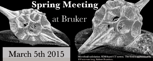
Conventional optical or electron microscopy allows visualizing two-dimensional images of a specimen surface or thin slices. However, in most cases a conclusion about original three-dimensional object structures cannot be made on the basis of two-dimensional information. X-ray computed tomography (micro-CT and nano-CT) is a non-invasive, 3-dimensional imaging technique that can be used to visualize internal structures of an object. Micro-CT offers researchers the ability to study the exterior and internal structure of tissues, whole organisms and soft and dense materials non-destructively. This talk will provide an overview of the method, discuss its applications, and show 3D videos that can be generated using microCT.
Bio
Ali Bahadur is an Applications Scientist with the Preclinical Imaging division of Bruker Biospin. Bruker offers a wide range of preclinical imaging systems including MRI, microPET, microSPECT, microCT and Optical Imaging modalities. Mr. Bahadur has worked in the Preclinical Imaging field for over a decade encompassing both academia and industry, mainly focusing on the application and use of microCT in drug discovery and development. At present, he provides product training and application support to Bruker’s microCT users in the Americas.
Robert Brandom is the northeast US salesman in the Bruker NanoAnalytics group, which provides analytical accessories for SEM, TEM, FIB, and electron microprobe systems. These accessories include EDS, EBSD, WDS, SEM-XRF and SEM-CT. His background includes teaching ore microscopy, using an automated QEM*SEM for the mining industry, training SEM users, and developing a novel SEM for the semiconductor industry.
“Seeing how biology feels: Cell mechanics measurements from microscopy”, Maria Kilfoil, PhD.University of Massachusetts Amherst
Abstract:
In this talk I will describe methods my lab has developed to measure mechanical properties from microscopic motions, and how study the mechanical behavior of active material systems either inspired by the cell, comprised of biopolymers and the motors that operate on them; or the cell itself. These systems can exhibit self-repair in response to their microenvironment, cooperatively build themselves, or move and do useful work to demonstrate their structures, rheological properties, and material patterns. Study of the mechanics of these behaviors is therefore valuable and requires new in situ approaches.
Bio:
Maria Kilfoil is an Assistant Professor of Physics at the University of Massachusetts Amherst. She received her BA from University of New Brunswick (Canada) in Physics, her PhD in Physics from Memorial University in Canada while doing her research in the laboratory of Professor Paul Callaghan in New Zealand, and then worked as a postdoctoral researcher at Harvard University in Physics and the Division of Engineering and Applied Science.
Her laboratory is focused on principles of active biological matter and understanding of biological (cellular) mechanics, making simple polymer-based models of the complex systems of cell-derived active materials. She has developed enabling microscopy-based microrheology and particle tracking techniques, and she is using these techniques to examine the motions and the mechanical properties of these complex systems. The specific systems she studies are two reconstituted, “bottom up” active systems (DNA/enzyme and cytoskeletal/enzyme), and a third, cellular system that exhibits aspects of both DNA and cytoskeletal active mechanics. While the goal of her research is to increase our understanding of biological (cellular) mechanics, in addition, active, soft, polymeric materials is an area in its nascence, so the chances of important industrial applications are currently without limitation.
Location
Bruker Optics Building
19 Fortune Drive
Billerica, MA 01821
Parking
Parking is unlimited, and is behind the Bruker Optics building.
Map
