
You are invited to the New England Society for Microscopy's 34th Annual Spring Workshops & Symposium at the Marine Biological Laboratory in Woods Hole, Massachusetts! The meeting will be held over two days - Thursday, April 27th, will feature two workshops which will allow a more in-depth exploration of state-of-the-art microscopy techniques. Registration and information about the Workshops can be found here. Friday, April 28th, will consist of a day-long Symposium featuring four technical talks, six 6-minute "lightning" talks, an exhibiting vendor session, and a buffet lunch.
Please, submit your lightning talk info here by April 21th. (Lightning talk speakers are entitled to free registration to this meeting).
Workshops include:
Intro to negative stain TEM
Sanchaita Das, University of Massachusetts Medical School, Worcester
&
Expansion Microscopy
Ruixuan Gao, Boyden Lab MIT
Register for either fantastic workshop by clicking here.
Schedule
|
Workshop Day April 27th
|
|
1:00 PM – Welcoming Remarks - Louis Kerr
|
|
1:10 PM – Workshops Part I
|
|
2:30 PM – Afternoon Coffee Break
|
|
3:00 PM – Workshops Part II
|
|
5:00 PM – Closing
|
|
Symposium Day April 28th
Meigs Room and Lobby
|
|
9:00 AM – Registration & Continental Breakfast
|
|
10:00 AM – Welcoming Remarks - NESM President
|
|
10:10 AM– Talk 1: Bioinspired Materials Mechanics: Traversing Size Scales; Alfred J. Crosby
|
|
10:50 AM – Lightning Talks
|
|
11:30 AM – Vendor Session
|
|
12:30 PM – Buffet lunch
|
|
1:45 PM – Talk 2: Imaging membrane remodeling at synapses; Avital Rodal
|
|
2:25 PM – Afternoon break
|
|
3:00 PM – Talk 3: Nanoscale Performance of Functional Materials and Active Systems; Bryan D. Huey
|
|
3:40 PM – Talk 4: Contribution of the electron microscope (and accessories) to Single-particle Cryo-electron microscopy, Method of the Year 2015; Steve Pfeiffer
|
|
4:20--Closing Remarks
|
Speaker Abstracts & Bios
Bioinspired Materials Mechanics: Traversing Size Scales
Alfred J. Crosby
Polymer Science & Engineering Department
University of Massachusetts Amherst
Abstract:
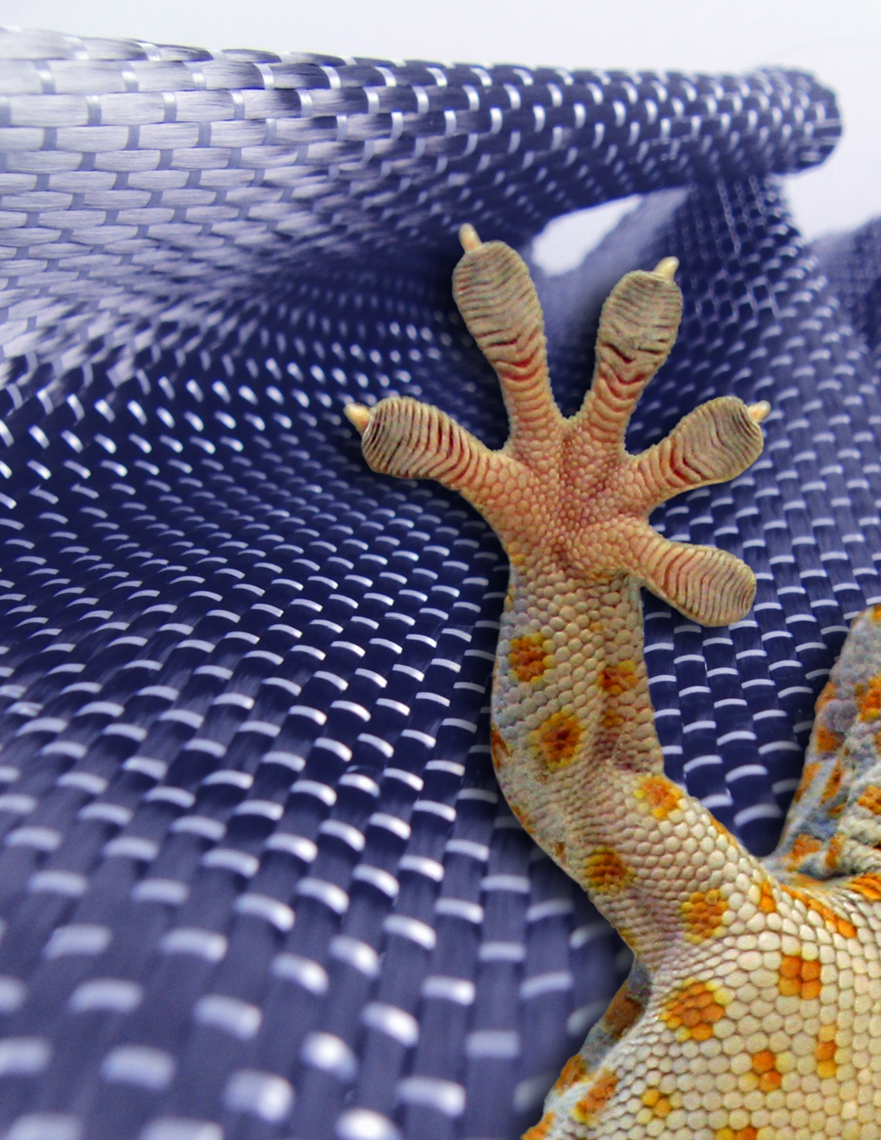
From geckos running along vertical surfaces at high speeds to mantis shrimp accelerating appendages at 10,000g, nature offers numerous, inspirational examples that use the control of materials mechanical properties to achieve impressive performances that are unmatched by current engineering capabilities. Our research group focuses on understanding the underlying principles of physics, chemistry, and engineering that govern such impressive performances and using them to develop synthetic materials systems that can have a positive impact on society. In this presentation, we highlight two examples: 1) the development of a gecko-inspired adhesive technology, called Geckskin®, and 2) the discovery of soft materials mesostructures, called mesoscale polymers, that offer new active control of mechanical properties and higher order assembly. Through these examples, we highlight how we rely upon the development of scaling principles and the use of microscopy to guide our understanding of natural systems and the development of new synthetic materials devices.
Bio:
Alfred J. Crosby is a Professor in the Polymer Science & Engineering Department at the University of Massachusetts Amherst and Co-Director of the Center for Evolutionary Materials. He has contributed significantly to the science and technology of soft materials, especially in the context of adhesion and bio-inspired materials. Prof. Crosby has received numerous awards and recognition for his research, including the National Science Foundation CAREER Award, the Army Research Office Young Investigator Award, the Adhesion Society Outstanding Young Scientist Award, the College of Natural Sciences Outstanding Research Award, Northwestern University’s Early Career Achievement Award in Materials Science and Engineering, and the University of Massachusetts Samuel F. Conti Faculty Fellowship Award. He has been recently elected to the National Academy of Inventors and as a Fellow of the American Physical Society, and his research has been covered extensively in the popular media, including Discovery Channel, Popular Science, CNET, NPR, Bloomberg Businessweek and CNN Money/Fortune Magazine, which named the Geckskin® project one of the Top 5 Science Breakthroughs of 2012. He is a co-founder of Felsuma, LLC, which is commercializing the Geckskin technology, and he serves as a consultant and advisor for several companies and professional organizations.
Links:
www.pse.umass.edu/acrosby
https://geckskin.umass.edu

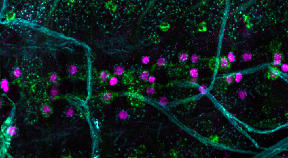
Central nervous system of a fruit fly larvae, labeled with the Alzheimer's Disease-implicated Amyloid Precursor Protein (green), neuronal nuclei (magenta) and neuronal membrane (cyan), and imaged by confocal microscopy.
Imaging membrane remodeling at synapses
Avital Rodal, PhD
Brandeis University
Abstract:
We are studying how highly specialized cells like neurons co-opt the machinery that traffics materials within and between cells to control their unique physiologies. We use live imaging in neurons of the fruit fly Drosophila melanogaster, together with in vitro studies of membrane remodeling proteins to understand the mechanisms by which neurons shape and sort intracellular membranes. We are also investigating how these processes go wrong in models of neurodegenerative disease, and testing if we can manipulate membrane traffic to rescue neurological defects.
Bio:
I am an Assistant Professor of Biology at Brandeis University. I was born in Ottawa, Canada, did my undergraduate studies at MIT, received my Ph.D. in Molecular and Cell Biology from the University of California, Berkeley in 2002. and did my post-doctoral work at the Massachusetts Institute of Technology. I am a Pew Scholar and recipient of the NIH New Innovator Award.
Nanoscale Performance of Functional Materials and Active Systems
Bryan D. Huey, PhD
Associate Professor and Director for Graduate Studies
Department of Materials Science & Engineering, UConn
Abstract:
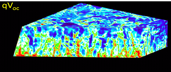
Nano- and micro- scale properties are crucial to the macroscopic performance of a wide range of functional and active materials and systems, such as many piezoelectrics, multiferroics, solar cells, and living biological cells. To directly investigate such effects at the nanoscale, we have developed High Speed, Tomographic, and other versions of Atomic Force Microscopy for unique insight into these diverse systems. With piezoelectrics, central to resonators, sensors, and ultrasonic actuators, geometric control is proven to locally enhance the converse-piezoelectric coefficient, with implications for a new class of transistors known as piezotronics (Keech et. al., Advanced Functional Materials, 2017). With ferroelectrics, movies of the domain switching process reveal unique nucleation and growth dynamics at grain boundaries and other microstructural defects (Huey et. al., J.A.Cer.S. cover article, 2012). Extending this approach to multiferroics, a stepwise polarization process was observed for the first time, which was then leveraged to develop the highest reported efficiency for a magnetoelectric switch (Heron et. al., Nature, 2014). For polycrystalline CdTe solar cells, novel photovoltaic performance maps reveal tremendous heterogeneity both at the surface, and as a function of depth (Luria et. al., Nature Energy, 2017). This suggests new pathways to improve their efficiency, which remains ~50% less than the theoretical limit yet already command ~5% of the world’s solar cell market. Finally, a novel combined AFM and 3d-fluorescence microscope allows simultaneous nanoscale probing of biological cells and direct detection of their living response. Macrophage cells assemble actin upon nanoNewton scale forces, while the loads applied by beating myocardial cells can be measured and their beat frequency even manipulated with AFM-based nano-CPR. These 6 systems, both inorganic and biological, especially exemplify the advantages of employing complementary microscopic methods including probe microscopy, optics, electron, and ion microscopy to reveal new fundamental properties and engineer improved future devices.
Bio:.
Bryan Huey is a UTC Professor of Engineering Innovation at the University of Connecticut. He is also the past chair of the 1200 person strong Basic Science Division of the American Ceramic Society. He is an expert in the development and application of advanced variations of Atomic Force Microscopy for studying piezoelectrics, multiferroics, photovoltaics, MEMS, and biological cells and tissue.
http://hueyafmlabs.mse.uconn.edu/


Micrograph- from this to, to a final product. Mm-Cpn with 120kV LaB6 @ 10Å resolution
Contribution of the electron microscope (and accessories) to Single-particle Cryo-electron microscopy, Method of the Year 2015
Steve Pfeiffer
Sales manager at Direct Electron, LP
Abstract:
“Nature Methods” selected single-particle cryo-electron microscopy as their choice for Method of the Year 2015. Single particle cryo-electron microscopy can be thought of as a three step process - a biochemical process of purification and physical specimen preparation, followed by imaging in an electron microscope and finally extensive image processing. This talk will focus primarily on the imaging portion of the process, discussing advances in microscopes, holders, phase plates, aberration correction and detectors.
The last few slides will address novel uses of direct detection devices in areas far removed from single particle cryo-electron microscopy, namely diffraction and in-situ TEM (materials).
Bio:
Steve received a Master’s degree in Biology, emphasis on Marine Invertebrate Zoology, from SDSU in 1982. There he learned EM on an RCA EMU-3H, cutting sections on an MT-1. Upon graduation he took a position with Max Cowan in the Developmental Neurobiology Lab at The Salk Institute. There he ran the electron microscope facility from 1982 to 1989, amongst other things developing double labelling techniques for EM. Leaving for greener pastures, he has worked at LifeCell (cryo-specimen prep for EM), BioRad & Noran (confocal microscopy), Carl Zeiss and Gatan (accessories for electron microscopy). Steve is currently at Direct Electron.
Location
Swope Conference Center
7 M B L Street
Woods Hole, MA
02543
Parking
Free parking facilities are located on-site. Upon arrival, please stop at the Swope Center to pick up a parking pass. Visitors will park at the Bar Neck lot.
Housing
