Join NESM for our annual Fall Symposium & Business Meeting on Thursday, November 17 at the Whitehead Institute in Cambridge, MA! The meeting will consist of four technical talks, a forty minute round of lightning talks, a coffee break, a buffet dinner, and a business meeting in which elections will be held for several positions on the Board of Directors. Election ballots will be distributed with the Fall E-Newsletter.
*The lightsheet workshop has been cancelled, we are sorry for the inconvenience.*
We are still accepting abstracts for one of six lightning talks.
To submit an abstract go to:
http://nesmicroscopy.org/lightning-talk-submission/
Meeting Schedule
12:45pm-Registration and Facility Tours
1:30pm-Welcome Remarks
1:40pm-Gabriella Kiss
2:20pm-Chen Xu
3:00pm-Coffee Break
3:30pm-Lightning Talks
4:10pm-Coffee Break
4:40pm-Andres Canales
5:20pm-Antonio Ambrosio
6:00pm-Buffet Dinner
7:00pm-Business Meeting
7:50pm-Closing remarks
Additional Speaker Information
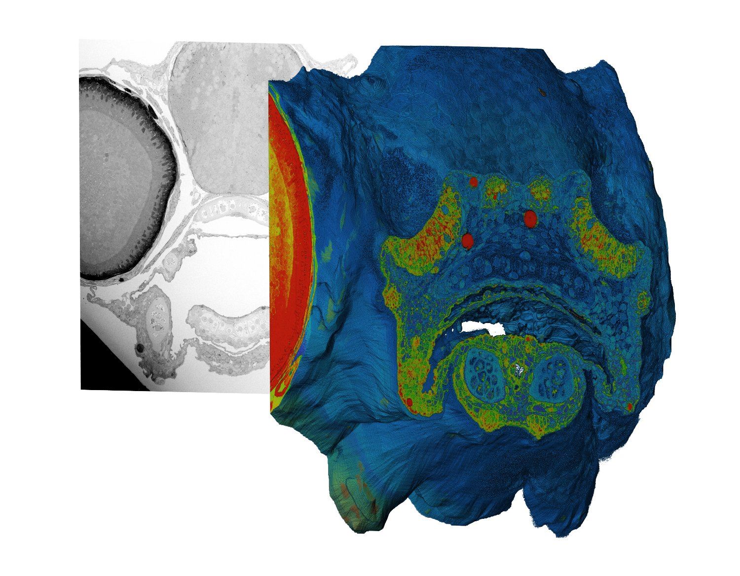
"3-Dimensional Characterization of Resin Embedded Biological Samples Using SBF-SEM and MED-SEM"
Gabriella Kiss, PhD Application Scientist FEI/ThermoFisher
Abstract:
3-dimensional characterization of resin embedded tissue samples in SEM or DualBeam FIB-SEM has demonstrated results equaling traditional 120 keV S/TEM tomography. With a continued interest in larger and larger tissue volumes, there has been a considerable advancement in SEM-based methods for imaging and 3-dimensional reconstruction in the past several years. Serial Block-Face SEM (SBF-SEM) involves combination of imaging and in-situ sectioning of resin embedded tissue within the SEM, allowing for automated imaging and subsequent reconstruction of volumes of tissue. To mitigate charge in what is generally a non-conductive specimen, low energy electrons or the use of low vacuum is employed. In 2014, FEI introduced and launched Teneo VolumeScope (VS), a SEM with integrated SBF and multi-energy deconvolution (MED-SEM). MED-SEM is a non-destructive technique that allows high-resolution imaging and reconstruction of the top layers of a sample. With multi-energy deconvolution one can overcome the traditional resolution limits set by mechanical slicing. After cutting a thin layer of the blockface using a diamond knife, freshly exposed tissue is imaged several times using various accelerating voltages. These images are subsequently used for deconvolving the information into several virtual subsurface layers. This cycle of physical and virtual sectioning offers isotropic datasets with excellent z-resolution and can be fully integrated and automated. This method will be presented as technique for isotropic 3-dimensional characterization of resin embedded tissue specimens.
Bio:
Gabriella completed her BSc and MSc in biotechnology, molecular biology and microbiology at University of Szeged in Hungary in 2007. She has started her PhD at University of Reading in UK in molecular virology in 2008. In the Neuman lab she studied enveloped virus structure, with her major focus on the role of the matrix protein in regulating virus assembly in influenza and coronaviruses. She used cryo-electron microscopy and single particle analysis for her projects. After finishing her postgraduate studies, she accepted a postdoctoral fellowship at Emory University in Atlanta USA in 2012. In the Wright lab she was studying different paramyxoviruses (RSV, Measles) using cryo-electron microscopy (cryo-EM) and cryo-electron tomography (cryo-ET). She joined FEI as an application scientist in 2014 where she learned SEM and FIB-SEM techniques and now she supports life science customers in a variety of electron microscopy applications.
"From CCD to DDD: A Story About TEM Recording Media"
Chen Xu, PhD Associate Professor and Cryo-EM Facility Director
Abstract:
Recent breakthroughs in the field of Cryo-EM largely benefit from the latest advances of digital cameras for TEM, especially direct electron detectors. Looking back, it has been quite a journey for TEM recording media’s development from film to high efficiency, high speed direct electron detecting devices. The talk tries to briefly review a history of recording medium for TEM electron imaging, and discuss some technical insights of late direct electron detectors. Some experience of using FEI Falcon II and Gatan K2 cameras is shared from a user perspective.
Bio:
Chen Xu had managed high-end Cryo-EM facility at Brandeis University for 15 years and trained many users. He has joined UMass Medical School since February 2016 to direct the newly setup Cryo-EM Core Facility.
"Flexible Fiber Probes for Multimodal Interaction with Neural Circuits"
Andres Canales, Graduate Student Materials Science & Engineering MIT
Abstract:
Understanding of complex neural systems requires the simultaneous probing of multiple signal modalities in the brain, such as electrical activity and chemical stimuli. Furthermore, it is possible to incorporate light-sensitive ion channels into the membranes of neurons, providing an additional handle to manipulate neural activity. I will present a novel family of neural probes, fabricated using a highly scalable thermal drawing process, capable of simultaneous electrical, chemical, and optical interrogation of the brain, and demonstrate their utility both in the brain and in the spinal cord of mice.
Bio:
Andres Canales completed his MSc in materials science and engineering in MIT. Now a PhD candidate in the same department, his work focuses on the development of flexible neural probes.
"Exploring Photon-Less Optical Microscopy"
Antonio Ambrosio, PhD Harvard University Center for Nanoscale Systems
Abstract:
Recently, the unique capability of AFM as weak-forces detector on the sample surface have been integrated in optical microscopes were the light illuminating the sample is the main driver of the forces detected by the AFM. Local thermal expansion due to light absorption as well as optical forces originated by the material polarizability can be detected as forces on the AFM probe. Such near-field detection schemes allow imaging and spectroscopy from the visible to the Mid-IR with nanometric spatial resolution although they do not detect in fact any light and are then photon-less. I will show results from ongoing experiments on different materials.
Bio:
Antonio Ambrosio is Senior Scientist at the Center for Nanoscale Systms at Harvard University. He has worked on different microscopy techniques on polymers, Carbon Nanotubes, 2D materials and metasurfaces. He co-authored more than 50 publications in international peer reviewed journals. He is currently editorial board member for Scientific Reports, a Nature Publishing Journal. His current research is devoted to explore the possibility of optical nano-imaging by means of detecting photo-induced optical forces between a sharp probe and the sample surface.
Location
McGovern Auditorium
Whitehead Institute
9 Cambridge Center
Cambridge, MA 02142
Parking
The Whitehead Institute is in the heart of Kendall Square/MIT and is within easy access of the Red Line T and bus routes.
- Traffic can be tough in Kendall Square so, if you decide to drive, please check Google directions to allow sufficient time.
- The map at http://en.parkopedia.com/parking/kendall_sq_cambridge/ shows local parking options. Whitehead Institute is located at the intersection marked with the location pin and the metered street parking pin.
- Street parking is limited to two hours and Cambridge Police are diligent in enforcing that.