
You are invited to the New England Society for Microscopy's 34th Annual Spring Workshops at the Marine Biological Laboratory on Thursday, April 27th! Below, you can find our workshop lineup and the schedule for the day. Registration for the Symposium on Friday, April 28th can be found below.
Location: Loeb 263
Schedule
1:00 PM – Welcoming Remarks
1:10 PM – Workshops Part I
2:30 PM – Afternoon Coffee Break
3:00 PM – Workshops Part II
5:00 PM – Closing
Workshop Descriptions
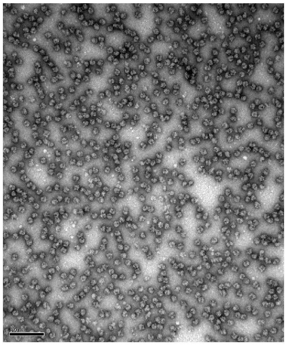
Intro to negative stain TEM
Sanchaita Das, University of Massachusetts Medical School, Worcester
Recent advances in Cryo-EM (Cryogenic Electron Microscopy) have brought about the resolution revolution in structural biology. However, Cryo-EM applications can be limited by protein size, flexibility, and imaging expense. Alternatively, Negative Stain Electron Microscopy (NS-EM) is a basic and efficient way of visualizing proteins that is not limited by a size barrier. Combined with other structural biology techniques like SAXS (Small Angle X-Ray Scattering) and NMR (Nuclear Magnetic Resonance), it can be a powerful tool for structure determination of small, flexible complexes. This method can also be utilized as an affordable screening technique for larger complexes being considered for Cryo-EM. This workshop will discuss the merits of NS-EM and demonstrate the procedure to see how simple, quick and highly informative the method can be.
Building: TBD, Marine Biological Laboratory
Wednesday Apr 27, 2017:
1:00 PM – Welcoming Remarks - Louis Kerr
1:10 PM – Workshops Part I : Discussion of NS-EM
2:30 PM – Afternoon Coffee Break
3:00 PM – Workshops Part II: Hands-on negative staining and imaging of purified ribosomes
5:00 PM – Closing remarks
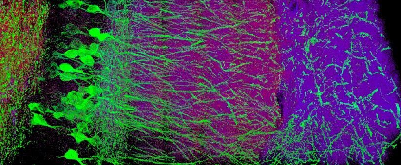
Expansion Microscopy
Ruixuan Gao, Boyden Lab MIT
http://syntheticneurobiology.org/
Expansion microscopy (ExM) allows for physical magnification of biological samples, including cells and tissues. In ExM, we link biomolecules and labels of interest within a sample, to a swellable polymer synthesized throughout the sample; we then mechanically homogenize the sample so as to enable isotropic expansion by several fold. Thus, ExM enables nanoscale imaging of large 3-D specimens, with a diversity of biomolecular species (e.g., proteins, RNA, etc) capable of identification and localization throughout extended samples. Since the technique can be carried out using simple chemicals (e.g., familiar antibodies and fluorescent proteins) and existing microscopy hardware, it is extremely practical to deploy in a variety of biological and medical settings.
In this short workshop, we will cover theory and practice of ExM, including underlying chemistry, sample preparation, optimized protocols for different tissue types and biomolecular species, as well as imaging of expanded samples and image processing.
Building: TBD, Marine Biological Laboratory
Wednesday Apr 27, 2017:
1:00 PM – Welcoming Remarks - Louis Kerr
1:10 PM – Workshops Part I : Discussion of ExM
2:30 PM – Afternoon Coffee Break
3:00 PM – Workshops Part II: Hands-on session
5:00 PM – Closing remarks